The History of Sleep Aids: From Ancient Herbal Remedies to Modern Precision Medicines

The History of Sleep Aids: From Ancient Herbal Remedies to Modern Precision Medicines

Thompson Le

Medical Affairs Intern

Humans have struggled with sleeplessness for thousands of years, long before we understood the biology of sleep. What has changed is how we approach insomnia, from experimenting with natural sedatives to developing targeted therapies based on neuroscience. The evolution of sleep aids reflects advances in chemistry, pharmacology, and patient safety.

Ancient Roots: Early Attempts to Induce Sleep

Long before modern medicine, cultures around the world used natural remedies to treat insomnia. Egyptians used opium-rich poppy extracts for sedation; Traditional Chinese Medicine relied on herbs like suan zao ren and valerian root, now known to influence GABAergic pathways. Greek and Roman healers mixed wine with plant alkaloids to promote sleep, while medieval Europeans used mandrake and belladonna, plants capable of sedation but also hallucinations and toxicity. These early treatments showed that sleep was recognized as vital, but pharmacologic attempts to induce it were often unpredictable and dangerous.

Poppy seeds. Center for Science in the Public Interest. Published September 11, 2024. https://www.cspi.org/poppy

Lustrea J. Valerian – Mother’s Little Helper During the Civil War. National Museum of Civil War Medicine. Published August 18, 2021. https://www.civilwarmed.org/valerian/

1800s–Early 1900s: The First Synthetic Sleep Medicines

The invention of chloral hydrate in 1869 marked the first major step toward modern sedatives. It provided more predictable sleep, though we now know it works by converting to trichloroethanol, a GABAergic compound. Bromide salts soon followed as sedatives and anxiolytics, but long-term use caused “bromism,” a toxic neurologic syndrome. These drugs were an improvement over herbal mixtures, yet their toxicity highlighted the need for safer options.

1900s–1950s: Barbiturates Dominate and Reveal Serious Risks

Barbiturates such as phenobarbital and secobarbital quickly became the standard for insomnia treatment. They were powerful and reliable but came with significant dangers: a narrow therapeutic window, high overdose risk, rapid tolerance, severe withdrawal, and deadly interactions with alcohol and opioids. Their widespread use underscored an emerging truth. Effective sedation without safety is not a sustainable long-term solution.

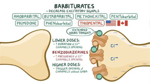
Anticonvulsants and anxiolytics: Barbiturates: Video. Osmosis. https://www.osmosis.org/learn/Anticonvulsants_and_anxiolytics:_Barbiturates

1960s–1980s: Benzodiazepines Offer a Safer Alternative

The introduction of benzodiazepines, starting with chlordiazepoxide (Librium) and diazepam (Valium), marked a turning point. These drugs enhanced GABA-A signaling with far lower overdose risk than barbiturates. Hypnotic benzodiazepines like flurazepam, temazepam, and triazolam became common for insomnia. However, over time, clinicians observed important limitations: tolerance, dependence, cognitive impairment, and rebound insomnia, especially with short-acting agents.

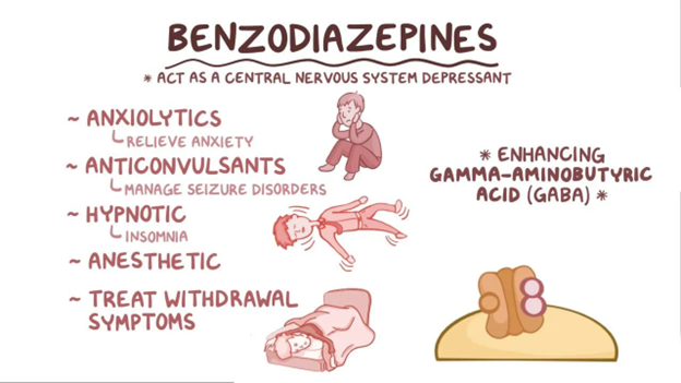
Anticonvulsants and anxiolytics: Benzodiazepines. Osmosis. https://www.osmosis.org/learn/Anticonvulsants_and_anxiolytics:_Benzodiazepines

1990s–2000s: Introduction of Non-Benzodiazepine “Z-Drugs”

Non-benzodiazepine hypnotics, including zolpidem, zaleplon, and eszopiclone, were developed to act at GABA-A receptors through a different binding profile than traditional benzodiazepines. These agents can improve sleep onset or maintenance depending on the molecule and formulation. However, Z-drugs are known to cause rebound insomnia when discontinued, particularly after higher doses or prolonged use. They also carry a Boxed Warning for complex sleep behaviors such as sleep-walking, sleep-driving, and other activities performed while not fully awake. The FDA later applied warnings about complex sleep behaviors across the broader sedative-hypnotic class after recognizing this adverse effect pattern. Misuse and abuse have also been reported in some populations, underscoring the need for appropriate prescribing and monitoring.

2010s–Present: Therapies Targeting the Orexin Pathway

Advances in sleep-wake neurobiology have contributed to the development of insomnia therapies with different mechanisms of action. One such approach involves Dual Orexin Receptor Antagonists (DORAs), including suvorexant, lemborexant, and daridorexant, which act by blocking wake-promoting orexin signaling. Clinical studies show that DORAs can improve sleep onset and maintenance without producing clinically meaningful rebound insomnia or withdrawal. However, like other Schedule IV hypnotics, they carry the potential for abuse and misuse, and they are associated with specific adverse effects such as sleep paralysis and hypnagogic/hypnopompic hallucinations. DORAs are also contraindicated in patients with narcolepsy, and individual agents may differ in next-day effects; for example, some data suggest daridorexant may have comparatively higher rates of next-day somnolence in certain populations.

Where We Are Today

Current guidelines emphasize Cognitive Behavioral Therapy for Insomnia (CBT-I) as first-line therapy, with medications used selectively and often short-term. Still, pharmacologic sleep aids remain important tools when guided by knowledge of mechanism, safety, and patient-specific factors.

Onward to More Personalized, Physiologic Sleep Therapies

From herbal potions to synthetic sedatives, benzodiazepines, Z-drugs, and orexin antagonists, each era of sleep aids built on and corrected the limitations of the last. Today, progress in circadian biology, pharmacogenomics, and neurophysiology is shaping the next generation of sleep treatments. The future of insomnia therapy will focus increasingly on understanding natural sleep patterns and developing more personalized approaches that support healthy, restorative sleep.

References

  1. López-Muñoz F, Ucha-Udabe R, Alamo C. The history of barbiturates a century after their clinical introduction. Neuropsychiatr Dis Treat. 2005;1(4):329-343.
  2. Neubauer DN. The evolution and development of insomnia pharmacotherapies. J Clin Sleep Med. 2007;3(5 Suppl):S11-S15.
  3. Edinoff AN, Wu N, Ghaffar YT, et al. Zolpidem: efficacy and side effects for insomnia. Health Psychol Res. 2021;9(1):24927. doi:10.52965/001c.24927
  4. Schoedel KA, Sun H, Sellers EM, et al. Assessment of the abuse potential of the orexin receptor antagonist suvorexant compared with zolpidem in a randomized crossover study. J Clin Psychopharmacol. 2016;36(4):314-323. doi:10.1097/JCP.0000000000000516
  5. Herring WJ, Connor KM, Snyder E, et al. Suvorexant in patients with insomnia: pooled analyses of three-month data from phase 3 randomized controlled clinical trials. J Clin Sleep Med. 2016;12(9):1215-1225. doi:10.5664/jcsm.6116
  6. Sateia MJ, Buysse DJ, Krystal AD, Neubauer DN, Heald JL. Clinical practice guideline for the pharmacologic treatment of chronic insomnia in adults: an American Academy of Sleep Medicine clinical practice guideline. J Clin Sleep Med. 2017;13(2):307-349. doi:10.5664/jcsm.6470
  7. Hu Z, Oh S, Ha TW, Hong JT, Oh KW. Sleep-aids derived from natural products. Biomol Ther (Seoul). 2018;26(4):343-349. doi:10.4062/biomolther.2018.099

The information provided is for general informational and educational purposes only. It is not intended to serve as medical advice, diagnosis, or treatment. The content is written by a licensed pharmacist and reflects general knowledge and expertise in the healthcare field, but it is not a substitute for professional medical advice from a qualified healthcare provider.

Always consult your physician, pharmacist, or other qualified healthcare professional before starting, stopping, or modifying any medication, treatment, or health regimen. Individual health conditions and needs vary, and only a healthcare professional can provide personalized advice tailored to your specific situation.

While we strive to ensure the accuracy and currency of the information presented, medical knowledge is constantly evolving, and errors or omissions may occur. The blog’s content does not cover all possible uses, precautions, side effects, or interactions of medications or treatments. Reliance on any information provided is solely at your own risk.

Links to external websites or resources are provided for convenience and do not imply endorsement or responsibility for their content.

Breaking Points: How OA and RA Challenge the Body in Different Ways

Breaking Points: How OA and RA Challenge the Body in Different Ways

Kameron Hicks

Medical Affairs Intern

Arthritis encompasses a group of inflammatory conditions that affect the joints and surrounding tissues, leading to pain, swelling, and limited movement. The two most common forms are osteoarthritis (OA) and rheumatoid arthritis (RA).¹

RA is an autoimmune disorder in which the body’s immune system attacks its own joint tissues, causing inflammation that leads to pain, stiffness, swelling, and redness, ultimately impairing joint mobility and function. Notably, RA symptoms often worsen after periods of rest and improve with activity as joint movement helps reduce inflammation and stiffness. The etiology of RA is believed to involve both genetic and environmental factors. Various alleles sharing a common amino acid sequence have been linked to RA development, and polymorphisms in other genes are associated with more severe disease. Cigarette smoking is the strongest environmental risk factor, while others include exposure to silica, asbestos, textile dust, and P. gingivalis. External antigens can trigger an autoimmune response that damages the joints.²

In contrast, osteoarthritis (OA) is a degenerative joint disease characterized by the progressive deterioration of articular cartilage. The loss of cartilage reduces cushioning and lubrication between bones, leading to friction, pain, stiffness, and decreased mobility. In contrast to RA, OA symptoms typically worsen with prolonged use and improve with rest, as mechanical stress aggravates joint pain. Pro-inflammatory mediators and proteolytic enzymes further contribute to joint destruction. As cartilage erosion progresses, chondrocytes proliferate and cluster, resulting in cartilage outgrowths and bone spurs. Continued damage to the collagen matrix triggers chondrocyte apoptosis, while abnormal collagen mineralization promotes subchondral bone thickening. Synovial inflammation and injury to surrounding joint structures, including ligaments, joint capsules, and menisci, may also occur.⁴

OA is classified as either primary or secondary. Primary OA develops with age in the absence of prior trauma or underlying disease, whereas secondary OA arises from joint abnormalities or injury. Major risk factors include advanced age, female sex, obesity, and a history of joint injury.⁵

Both OA and RA are characterized by joint pain, swelling, and reduced mobility. Although their specific causes are not fully understood, a combination of genetic, environmental, and lifestyle factors contributes to their development.

References:

  1. Johns Hopkins Medicine. Arthritis. Updated February 2025. Accessed October 14, 2025. https://www.hopkinsmedicine.org/health/conditions-and-diseases/arthritis
  2. American College of Rheumatology. Rheumatoid Arthritis. Updated February 2025. Accessed October 14, 2025. https://rheumatology.org/patients/rheumatoid-arthritis
  3. Chauhan K, Jandu JS, Brent LH, et al. Rheumatoid Arthritis. [Updated 2023 May 25]. In: StatPearls [Internet]. Treasure Island (FL): StatPearls Publishing; 2025 Jan-. https://www.ncbi.nlm.nih.gov/books/NBK441999/
  4. Cleveland Clinic. Osteoarthritis: Symptoms, Causes & Treatment Options. Updated October 2, 2023. Accessed October 14, 2025. https://my.clevelandclinic.org/health/diseases/5599-osteoarthritis
  5. Sen R, Hurley JA. Osteoarthritis. [Updated 2023 Feb 20]. In: StatPearls [Internet]. Treasure Island (FL): StatPearls Publishing; 2025 Jan-. https://www.ncbi.nlm.nih.gov/books/NBK482326/
  6. Osteoarthritis vs. Rheumatoid Arthritis. MedComic. Accessed October 14, 2025. https://www.medcomic.com/medcomic/osteoarthritis-vs-rheumatoid-arthritis

The information provided is for general informational and educational purposes only. It is not intended to serve as medical advice, diagnosis, or treatment. The content is written by a licensed pharmacist and reflects general knowledge and expertise in the healthcare field, but it is not a substitute for professional medical advice from a qualified healthcare provider.

Always consult your physician, pharmacist, or other qualified healthcare professional before starting, stopping, or modifying any medication, treatment, or health regimen. Individual health conditions and needs vary, and only a healthcare professional can provide personalized advice tailored to your specific situation.

While we strive to ensure the accuracy and currency of the information presented, medical knowledge is constantly evolving, and errors or omissions may occur. The blog’s content does not cover all possible uses, precautions, side effects, or interactions of medications or treatments. Reliance on any information provided is solely at your own risk.

Links to external websites or resources are provided for convenience and do not imply endorsement or responsibility for their content.

Trending Research and Innovation in Pain Management

Trending Research and Innovation in Pain Management

Logan Anderson, Pharm.D., RPh

Galt Pharmaceuticals Medical Affairs Fellow

Emerging Trends

In a study of 90,769 US adults, chronic pain increased by 18% and high-impact chronic pain (HICP) increased by 13% compared to pre- COVID-19 pandemic reports (2019-2023). Chronic pain was described as pain occurring at least every other day for 3 or more months while HICP was described as chronic pain that limits activities at least every other day. Long COVID was found to explain roughly 13% of the increase in all chronic pain from 2019 to 2023. Other potential contributors include increases in the prevalence of obesity, depressive symptoms, and anxiety as well as increased demand on healthcare institutions affecting treatment outcomes.1

Research Advancement

Sphingosine 1-Phosphate (S1P) receptor antagonists (i.e., ozanimod and ertrasimod) prevent lymphocytes from leaving lymph nodes en route to the central nervous system and intestine. This mechanism has been found useful for the treatment of multiple sclerosis and ulcerative colitis.2,3 A recent study by Ginacotti et al. has demonstrated that S1P receptor 1 activation contributes to neuropathic pain. Administration of ozanimod into the rostral ventral medulla – a key structure of descending pain modulation – reduced behavioral hypersensitivities by incorporating noradrenergic and serotonergic inhibitory pathways.4 This study encourages further research into the use of S1P receptor antagonists for the treatment of neuropathic pain.

Novel Targets

Cortical astrocytes were identified as a potential target for migraine treatment. Bree et al. evaluated the effect of activating visual cortex astrocytes in rats. They observed migraine-like behaviors and prolonged meningeal nociceptor discharge which was capable of being blocked by inhibiting CGRP signaling.5

New Approvals

Journavx (suzetrigine) was recently approved in January of this year as the first sodium channel blocker for the treatment of moderate to severe acute pain in adults.6 Suzetrigine selectively inhibits Nav 1.8 voltage-gated sodium channels to block ascending pain signals from peripheral sensory neurons.7 The Institute for Clinical and Economic Review (ICER) assessed the effectiveness and value of suzetrigine in February. The ICER panel of experts found that there was adequate evidence for the benefit of suzetrigine over non-systemic therapies but determined that there was not adequate evidence for the benefit of suzetrigine over opioids or oral NSAIDs in addition to non-systemic therapies. The ICER panel was divided on the valuation of suzetrigine. The majority of panelists stated that suzetrigine had intermediate long term value with the lack of long-term safety data and ambiguous phase III results being a point of discussion.8

References

  1. Zajacova, Annaa,*; Grol-Prokopczyk, Hannab; Nahin, Richard L.c. Pain among US adults before, during, and after the COVID-19 pandemic: a study using the 2019 to 2023 National Health Interview Survey. PAIN ():10.1097/j.pain.0000000000003764, August 5, 2025. | DOI: 10.1097/j.pain.0000000000003764
  2. Lexi-Drugs. UpToDate Lexidrug. UpToDate Inc. https://online.lexi.com. Accessed August 25, 2025.
  3. Lexi-Drugs. UpToDate Lexidrug. UpToDate Inc. https://online.lexi.com. Accessed August 25, 2025.
  4. Giancotti, Luigino Antonio; Squillace, Silvia; Chen, Zhomou; Lauro, Filomena; Li, Ying; Salvemini, Daniela*. Ozanimod, a functional sphingosine-1-phosphate receptor 1 antagonist, restores brainstem descending pain pathways in murine models of neuropathic pain. PAIN ():10.1097/j.pain.0000000000003770, August 4, 2025. | DOI: 10.1097/j.pain.0000000000003770
  5. Bree, Dara; Zhao, Jun; Stratton, Jennifer; Levy, Dan*. Cortical astrocyte activation triggers meningeal nociception and migraine-like pain. PAIN ():10.1097/j.pain.0000000000003737, July 30, 2025. | DOI: 10.1097/j.pain.0000000000003737
  6. FDA Approves Novel Non-Opioid Treatment for Moderate to Severe Acute Pain. U.S. Food and Drug Administration. Published January 30, 2025. https://www.fda.gov/news-events/press-announcements/fda-approves-novel-non-opioid-treatment-moderate-severe-acute-pain
  7. Lexi-Drugs. UpToDate Lexidrug. UpToDate Inc. https://online.lexi.com. Accessed August 25, 2025.
  8. Nikitin D, Rind DM, McQueen B, et al. The effectiveness and value of suzetrigine for moderate to severe acute pain: A summary from the Institute for Clinical and Economic Review’s Midwest Comparative Effectiveness Public Advisory Council. Journal of Managed Care & Specialty Pharmacy. 2025;31(7):729-734. doi:https://doi.org/10.18553/jmcp.2025.31.7.729

The information provided is for general informational and educational purposes only. It is not intended to serve as medical advice, diagnosis, or treatment. The content is written by a licensed pharmacist and reflects general knowledge and expertise in the healthcare field, but it is not a substitute for professional medical advice from a qualified healthcare provider.

Always consult your physician, pharmacist, or other qualified healthcare professional before starting, stopping, or modifying any medication, treatment, or health regimen. Individual health conditions and needs vary, and only a healthcare professional can provide personalized advice tailored to your specific situation.

While we strive to ensure the accuracy and currency of the information presented, medical knowledge is constantly evolving, and errors or omissions may occur. The blog’s content does not cover all possible uses, precautions, side effects, or interactions of medications or treatments. Reliance on any information provided is solely at your own risk.

Links to external websites or resources are provided for convenience and do not imply endorsement or responsibility for their content.

How the Sleep Cycle Works: Stages, Duration, and Tips for Better Rest

How the Sleep Cycle Works: Stages, Duration, and Tips for Better Rest

Viral Patel
Viral Patel

Galt Pharmaceuticals Medical Affairs Fellow, 2024-2025

Humans cycle through 2 phases of sleep, rapid eye movement (REM) and nonrapid eye movement (NREM). NREM sleep can be further divided into 3 stages N1, N2, and N3. Each phase of sleep includes variations in muscle tone, brain wave patterns, and eye movements.1

  1. N1 (Stage 1 NREM)
    1. This is the lightest stage of sleep, representing the transition from wakefulness to sleep. It is characterized by low-amplitude, mixed-frequency EEG activity, and slow rolling eye movements. Muscle tone decreases slightly.2
  2. N2 (Stage 2 NREM)
    1. This stage constitutes the majority of total sleep time. It is marked by the presence of sleep spindles and K-complexes on EEG. Eye movements stop and muscle tone is further reduced.2
  3. N3 (Stage 3 NREM)
    1. This is the deepest stage of NREM sleep, characterized by high-amplitude and low-frequency delta waves. It is associated with the highest arousal threshold and is considered restorative sleep.2
  4. REM Sleep
    1. REM is characterized by rapid eye movements, low-amplitude mixed-frequency EEG (similar to wakefulness), and muscle atonia. This stage is associated with vivid dreaming and increased brain activity.3

The body cycles through all stages approximately 4 to 6 times each night, averaging 90-120 minutes per cycle.1 N1 ranges from 1-7 minutes at the start of the night and is often shorter in subsequent cycles. N2 is approximately 10-25 minutes in the first cycle and will increase in duration in later cycles. N3 takes 20-40 minutes in the first cycle, decreasing in duration in later cycles. REM sleep typically lasts 10 minutes in the first cycle and will increase to 30-40 minutes by the last cycle.4

There are many ways to improve sleep quality:

  • Prioritizing adequate sleep duration
  • Maintaining regular sleep routines
  • Optimizing the sleep environment
  • Minimizing evening stimulants
  • Addressing comorbid sleep disorders

These strategies can help improve sleep quality and preserve the integrity of NREM and REM sleep cycles.5

References

  1. Patel AK, Reddy V, Shumway KR, et al. Physiology, Sleep Stages. [Updated 2024 Jan 26]. In: StatPearls [Internet]. Treasure Island (FL): StatPearls Publishing; 2025 Jan-. Available from: https://www.ncbi.nlm.nih.gov/books/NBK526132/
  2. Chokroverty S. Overview of sleep & sleep disorders. Indian J Med Res. 2010;131:126-140.
  3. Rodenbeck A. Biologische Grundlagen des Schlafens und Wachens [Biological principles of sleep and wake]. Bundesgesundheitsblatt Gesundheitsforschung Gesundheitsschutz. 2011;54(12):1270-1275. doi:10.1007/s00103-011-1373-3
  4. Gottesman RF, Lutsey PL, Benveniste H, et al. Impact of Sleep Disorders and Disturbed Sleep on Brain Health: A Scientific Statement From the American Heart Association. Stroke. 2024;55(3):e61-e76. doi:10.1161/STR.0000000000000453
  5. Baranwal N, Yu PK, Siegel NS. Sleep physiology, pathophysiology, and sleep hygiene. Prog Cardiovasc Dis. 2023;77:59-69. doi:10.1016/j.pcad.2023.02.005

The information provided is for general informational and educational purposes only. It is not intended to serve as medical advice, diagnosis, or treatment. The content is written by a licensed pharmacist and reflects general knowledge and expertise in the healthcare field, but it is not a substitute for professional medical advice from a qualified healthcare provider.

Always consult your physician, pharmacist, or other qualified healthcare professional before starting, stopping, or modifying any medication, treatment, or health regimen. Individual health conditions and needs vary, and only a healthcare professional can provide personalized advice tailored to your specific situation.

While we strive to ensure the accuracy and currency of the information presented, medical knowledge is constantly evolving, and errors or omissions may occur. The blog’s content does not cover all possible uses, precautions, side effects, or interactions of medications or treatments. Reliance on any information provided is solely at your own risk.

Links to external websites or resources are provided for convenience and do not imply endorsement or responsibility for their content.

The Role of NSAIDs in Managing Inflammatory Conditions

body with joints highlighted

The Role of NSAIDs in Managing Inflammatory Conditions

Viral Patel
Viral Patel

Galt Pharmaceuticals Medical Affairs Fellow, 2024-2025

Nonsteroidal anti-inflammatory drugs (NSAIDs) are a class of medication used to treat various inflammatory conditions due to their analgesic, antipyretic, and anti-inflammatory properties. NSAIDs main mechanism of action is the inhibition of the enzyme cyclooxygenase (COX). This enzyme is crucial for the synthesis of prostaglandins which are involved in various inflammatory processes.1

Key inflammatory conditions treated with NSAIDs include:

  1. Rheumatoid Arthritis (RA)
    1. Rheumatoid arthritis is a systemic autoimmune disease characterized by inflammatory arthritis and extra-articular involvement. It is a chronic inflammatory disorder caused in many cases by the interaction between genes and environmental factors.2
    2. NSAIDs are commonly used to manage pain and inflammation in RA. They provide symptomatic relief but do not alter the disease course.3
  1. Osteoarthritis (OA)
    1. Osteoarthritis is the most common form of arthritis in the world. OA is degenerative joint disease characterized by the breakdown of cartilage and the underlying bone, causing pain, stiffness, and limited movement.4
    2. NSAIDs are effective in reducing pain and improving function in patients with OA.
  1. Acute gout
    1. Gout is one of the most common causes of chronic inflammatory arthritis in the United States. Gout is characterized by monohydrate crystals deposited in tissue.5
    2. NSAIDs are first-line treatment for acute gout flares, providing significant pain relief and reduction in inflammation in affected joints.6
  1. Ankylosing Spondylitis (AS)
    1. Ankylosing spondylitis is a chronic, inflammatory disease of the axial spine. Chronic back pain and progressive spinal stiffness are the most common features of this disease.7
    2. NSAIDs are recommended as first-line therapy for reducing pain and stiffness in AS. Continuous NSAID treatment is often advised for persistently active disease per the American College of Rheumatology.8
  1. Primary dysmenorrhea
    1. Primary dysmenorrhea is defined as cramping pain in the lower abdomen occurring just before or during menstruation, in the absence of other diseases.9
    2. NSAIDs have been shown to be effective agents in managing pain associated with primary dysmenorrhea.10

The dosage and duration of NSAID therapy should be tailored to the individual patient’s needs, balancing efficacy with the risk of adverse effects, particularly gastrointestinal, renal and cardiovascular complications.

References

  1. Ghlichloo I, Gerriets V. Nonsteroidal Anti-Inflammatory Drugs (NSAIDs) [Updated 2023 May 1]. In: StatPearls [Internet]. Treasure Island (FL): StatPearls Publishing; 2025 Jan-. Available from: https://www.ncbi.nlm.nih.gov/books/NBK547742/
  2. Chauhan K, Jandu JS, Brent LH, et al. Rheumatoid Arthritis. [Updated 2023 May 25]. In: StatPearls [Internet]. Treasure Island (FL): StatPearls Publishing; 2025 Jan-. Available from: https://www.ncbi.nlm.nih.gov/books/NBK441999/
  3. Braun J, Baraliakos X, Westhoff T. Nonsteroidal anti-inflammatory drugs and cardiovascular risk – a matter of indication. Semin Arthritis Rheum. 2020;50(2):285-288. doi:10.1016/j.semarthrit.2019.07.012
  4. Sen R, Hurley JA. Osteoarthritis. [Updated 2023 Feb 20]. In: StatPearls [Internet]. Treasure Island (FL): StatPearls Publishing; 2025 Jan-. Available from: https://www.ncbi.nlm.nih.gov/books/NBK482326/
  5. Fenando A, Rednam M, Gujarathi R, et al. Gout. [Updated 2024 Feb 12]. In: StatPearls [Internet]. Treasure Island (FL): StatPearls Publishing; 2025 Jan-. Available from: https://www.ncbi.nlm.nih.gov/books/NBK546606/
  6. van Durme CM, Wechalekar MD, Landewé RB, et al. Non-steroidal anti-inflammatory drugs for acute gout. Cochrane Database Syst Rev. 2021;12(12):CD010120. Published 2021 Dec 9. doi:10.1002/14651858.CD010120.pub3
  7. Wenker KJ, Quint JM. Ankylosing Spondylitis. [Updated 2023 Jun 20]. In: StatPearls [Internet]. Treasure Island (FL): StatPearls Publishing; 2025 Jan-. Available from: https://www.ncbi.nlm.nih.gov/books/NBK470173/
  8. Poddubnyy D, van der Heijde D. Therapeutic controversies in spondyloarthritis: nonsteroidal anti-inflammatory drugs. Rheum Dis Clin North Am. 2012;38(3):601-611. doi:10.1016/j.rdc.2012.08.005
  9. Coco AS. Primary dysmenorrhea. Am Fam Physician. 1999;60(2):489-496.
  10. Itani R, Soubra L, Karout S, Rahme D, Karout L, Khojah HMJ. Primary Dysmenorrhea: Pathophysiology, Diagnosis, and Treatment Updates. Korean J Fam Med. 2022;43(2):101-108. doi:10.4082/kjfm.21.0103

The information provided is for general informational and educational purposes only. It is not intended to serve as medical advice, diagnosis, or treatment. The content is written by a licensed pharmacist and reflects general knowledge and expertise in the healthcare field, but it is not a substitute for professional medical advice from a qualified healthcare provider.

Always consult your physician, pharmacist, or other qualified healthcare professional before starting, stopping, or modifying any medication, treatment, or health regimen. Individual health conditions and needs vary, and only a healthcare professional can provide personalized advice tailored to your specific situation.

While we strive to ensure the accuracy and currency of the information presented, medical knowledge is constantly evolving, and errors or omissions may occur. The blog’s content does not cover all possible uses, precautions, side effects, or interactions of medications or treatments. Reliance on any information provided is solely at your own risk.

Links to external websites or resources are provided for convenience and do not imply endorsement or responsibility for their content.

Oral Hygiene Practices for Preventing Oral Thrush

Oral hygiene background

Oral Hygiene Practices for Preventing Oral Thrush

Scott Chappell
Scott Chappell

Galt Pharmaceuticals Director of Medical Affairs

Good oral hygiene practices are essential in preventing oral thrush, also known as oral candidiasis, which is primarily caused by the overgrowth of Candida species, particularly Candida albicans. Here are some evidence-based strategies to maintain good oral hygiene and prevent oral thrush.

  1. Regular Brushing and Flossing: Brush teeth at least twice daily with fluoride toothpaste and floss daily to remove dental plaque, which can harbor Candida. The American Dental Association emphasizes the importance of effective plaque removal and regular dental visits to promote oral health.[1]

  2. Use of Antimicrobial Mouthwashes: Mouthwashes containing chlorhexidine digluconate or cetylpyridinium chloride have been shown to impair the virulent traits of Candida albicans and other oral pathogens, thus helping to maintain microbial homeostasis in the oral cavity. However, chlorhexidine should be used with caution due to potential side effects.[2]

  3. Maintain Salivary Flow: Adequate salivary flow is crucial as saliva has antifungal properties. Patients with dry mouth (xerostomia) should use mechanical salivary stimulants, oral moisturizers, or systemic medications to promote saliva to manage dry mouth and reduce the risk of oral candidiasis. The American Dental Association recommends these measures for managing xerostomia and preventing candidiasis.[3]

  4. Proper Denture Hygiene: For denture wearers, it is important to clean dentures daily and remove them at night. Disinfecting dentures with solutions such as 2% chlorhexidine gluconate can prevent reinfection. Denture stomatitis, a form of oral candidiasis, is often associated with poor denture hygiene. [4-5]

  5. Avoiding Risk Factors: Minimize the use of broad-spectrum antibiotics and corticosteroids, which can disrupt the normal oral microbiota and promote fungal overgrowth. Patients should also avoid high-sugar diets and smoking, as these can increase the risk of oral candidiasis.[6]

  6. Regular Dental Check-ups: Regular dental visits are essential for early detection and management of oral conditions that may predispose to candidiasis. Dentists can provide professional cleaning and tailored advice on maintaining oral hygiene.[1]

By adhering to these practices, individuals can significantly reduce their risk of developing oral thrush and maintain overall oral health.

References:

  1. Managing Xerostomia and Salivary Gland Hypofunction: Executive Summary of a Report From the American Dental Association Council on Scientific Affairs. Plemons JM, Al-Hashimi I, Marek CL. Journal of the American Dental Association (1939). 2014;145(8):867-73. doi:10.14219/jada.2014.44.
  2. In Vitro Effects of Commercial Mouthwashes on Several Virulence Traits of Candida Albicans, Viridans Streptococci and Enterococcus Faecalis Colonizing the Oral Cavity. Ardizzoni A, Pericolini E, Paulone S, et al. PloS One. 2018;13(11):e0207262. doi:10.1371/journal.pone.0207262.
  3. Common Oral Conditions: A Review.Stoopler ET, Villa A, Bindakhil M, Díaz DLO, Sollecito TP. Jama. 2024;331(12):1045-1054. doi:10.1001/jama.2024.0953.
  4. Oral Fungal Infections. Muzyka BC. Dental Clinics of North America. 2005;49(1):49-65, viii. doi:10.1016/j.cden.2004.07.007.
  5. Management of Chronic Atrophic Candidiasis (Denture Stomatitis)-a Narrative Review. Abuhajar E, Ali K, Zulfiqar G, et al. International Journal of Environmental Research and Public Health. 2023;20(4):3029. doi:10.3390/ijerph20043029.
  6. Oral Candidiasis. Akpan A, Morgan R. Postgraduate Medical Journal. 2002;78(922):455-9. doi:10.1136/pmj.78.922.455.

The information provided is for general informational and educational purposes only. It is not intended to serve as medical advice, diagnosis, or treatment. The content is written by a licensed pharmacist and reflects general knowledge and expertise in the healthcare field, but it is not a substitute for professional medical advice from a qualified healthcare provider.

Always consult your physician, pharmacist, or other qualified healthcare professional before starting, stopping, or modifying any medication, treatment, or health regimen. Individual health conditions and needs vary, and only a healthcare professional can provide personalized advice tailored to your specific situation.

While we strive to ensure the accuracy and currency of the information presented, medical knowledge is constantly evolving, and errors or omissions may occur. The blog’s content does not cover all possible uses, precautions, side effects, or interactions of medications or treatments. Reliance on any information provided is solely at your own risk.

Links to external websites or resources are provided for convenience and do not imply endorsement or responsibility for their content.


DSM 5 and Insomnia Basics

DSM 5 and Insomnia Basics

Viral Patel
Viral Patel

Galt Pharmaceuticals Medical Affairs Fellow, 2024-2025

What is Insomnia?

Insomnia is characterized by dissatisfaction with sleep quantity/quality. It is associated with difficulty initiating sleep, maintaining sleep, or early-morning awakenings. These symptoms must occur for at least three nights per week and be present for at least the last three months. Untreated insomnia can cause significant distress or impairment in social, occupational, or other important areas of life.3

DSM 5 Characteristics of Insomnia

The Diagnostic and Statistical Manual of Mental Illnesses (DSM-5) provides clear and highly detailed definitions of mental health and brain related problems.1 In regard to insomnia, the following list highlights the diagnostic criteria utilized in the guideline:

  1. A predominant complaint of dissatisfaction with sleep quantity or quality, associated with one (or more) of the following symptoms:
    • Difficulty initiating sleep.
      1. In children, this may manifest as difficulty initiating sleep without caregiver intervention.
    • Difficulty maintaining sleep, characterized by frequent awakenings or problems returning to sleep after awakenings.
      1. In children, this may manifest as difficulty returning to sleep without caregiver intervention.
    • Early morning awakening with inability to return to sleep.
  2. Sleep disturbance causes clinically significant distress or impairment in social, occupational, educational, academic, behavioral, or other important areas of functioning.
  3. Sleep difficulty occurs at least 3 nights per week.
  4. Sleep difficulty is present for at least 3 months.
  5. Sleep difficulty occurs despite adequate opportunity for sleep.
  6. The insomnia is not better explained by and does not occur exclusively during the course of another sleep-wake disorder (e.g., narcolepsy, a breathing-related sleep disorder, a circadian rhythm sleep-wake disorder, parasomnia).
  7. The insomnia is not attributable to the physiological effects of a substance (e.g., a drug of abuse, a medication).
  8. Coexisting mental disorders and medical conditions do not adequately explain the predominant complaint of insomnia.2

Treatment for Insomnia

Treatment for insomnia disorder typically includes cognitive-behavioral therapy (CBT) as the first-line treatment. Pharmacotherapy may be considered when CBT is insufficient or inaccessible.2

References

  1. Cleveland Clinic. DSM-5. Published October 14, 2022. https://my.clevelandclinic.org/health/articles/24291-diagnostic-and-statistical-manual-dsm-5
  2. Sleep-Wake Disorders. Diagnostic and Statistical Manual of Mental Disorders. March 2025. doi:10.1176/appi.books.9780890425787.x12_Sleep-Wake_Disorders
  3. Winkelman JW. Clinical practice. Insomnia disorder. N Engl J Med. (2015) 373(15):1437–44. doi: 10.1056/NEJMcp1412740

The information provided is for general informational and educational purposes only. It is not intended to serve as medical advice, diagnosis, or treatment. The content is written by a licensed pharmacist and reflects general knowledge and expertise in the healthcare field, but it is not a substitute for professional medical advice from a qualified healthcare provider.

Always consult your physician, pharmacist, or other qualified healthcare professional before starting, stopping, or modifying any medication, treatment, or health regimen. Individual health conditions and needs vary, and only a healthcare professional can provide personalized advice tailored to your specific situation.

While we strive to ensure the accuracy and currency of the information presented, medical knowledge is constantly evolving, and errors or omissions may occur. The blog’s content does not cover all possible uses, precautions, side effects, or interactions of medications or treatments. Reliance on any information provided is solely at your own risk.

Links to external websites or resources are provided for convenience and do not imply endorsement or responsibility for their content.


Managing Breakthrough Pain in Rheumatoid Arthritis

Managing Breakthrough Pain in Rheumatoid Arthritis

Scott Chappell
Scott Chappell

Galt Pharmaceuticals Director of Medical Affairs

Adjusting current DMARD (Disease Modifying Antirheumatic Drugs) therapy, utilizing non-pharmacologic therapies and employing NSAIDs play significant roles in managing breakthrough symptoms for rheumatoid arthritis (RA) management.

Managing Breakthrough Pain with Current DMARD Therapy

Managing of breakthrough symptoms with current DMARD therapy, the treatment options are guided by the 2021 American College of Rheumatology (ACR) guidelines.[1]

  1. Optimization of Current DMARD Therapy: If a patient is experiencing breakthrough symptoms, the first step is to optimize the current disease-modifying antirheumatic drug (DMARD) regimen. This may involve increasing the dose of the current DMARD or adding another conventional synthetic DMARD (csDMARD) such as methotrexate, hydroxychloroquine, sulfasalazine, or leflunomide.[1]

  2. Switching or Adding Biologic DMARDs (bDMARDs): If optimization of csDMARDs is insufficient, the next step is to consider biologic DMARDs. Options include TNF inhibitors (e.g., etanercept, adalimumab, infliximab), IL-6 receptor inhibitors (e.g., tocilizumab, sarilumab), T cell costimulatory inhibitors (e.g., abatacept), and B cell depleting agents (e.g., rituximab). [1-2]

  3. Targeted Synthetic DMARDs (tsDMARDs): For patients who do not respond adequately to bDMARDs, targeted synthetic DMARDs such as Janus kinase (JAK) inhibitors (e.g., tofacitinib, baricitinib, Upadacitinib) can be considered.[1]

  4. Switching Mechanisms of Action: If a patient fails to respond to one class of bDMARDs, switching to a different mechanism of action (e.g., from a TNF inhibitor to an IL-6 receptor inhibitor or a JAK inhibitor) is recommended. [3-4]

  5. Combination Therapy: Combining csDMARDs with bDMARDs or tsDMARDs can be effective in managing breakthrough symptoms and achieving better disease control. [1-2]

  6. Glucocorticoids: Short-term use of glucocorticoids can be considered for rapid symptom relief, but long-term use is generally discouraged due to potential adverse effects.[1][5]

These strategies should be tailored to the individual patient, considering factors such as disease severity, comorbidities, and previous treatment responses. Regular monitoring and a treat-to-target approach are essential to optimize outcomes.[1][6]

Non-Pharmacologic Interventions for Breakthrough Pain

The 2022 American College of Rheumatology (ACR) guidelines recommend several non-pharmacologic interventions for RA management. [7] 

  1. Exercise Therapy: Regular aerobic and strength exercises improve physical function and reduce pain.

  2. Therapeutic Patient Education: Educating patients on self-management strategies, including joint protection and energy conservation, is beneficial.

  3. Physical and Occupational Therapy: Tailored exercise programs, assistive devices, and ergonomic advice help maintain joint function and reduce pain.

  4. Orthoses and Assistive Devices: Devices like wrist splints and foot orthoses provide joint support and pain relief.

  5. Dietary Interventions: Diets such as the Mediterranean diet, which emphasizes vegetables, fruits, whole grains, and healthy fats, may have anti-inflammatory effects.

  6. Cognitive Behavioral Therapy (CBT): CBT helps manage the psychological impact of RA, improving overall quality of life.

  7. Balneotherapy: Mineral baths can provide symptomatic relief.

  8. Mind-Body Approaches: Techniques like mindfulness, meditation, and biofeedback can help manage pain and stress.

NSAIDs for Breakthrough Pain

NSAIDs are effective for managing breakthrough pain in RA by reducing inflammation and pain. However, NSAIDs should be used at the lowest effective dose for the shortest duration to minimize risks such as gastrointestinal complications and cardiovascular events.[3]

In summary, adjusting DMARD therapy, utilizing non-pharmacologic therapies, and employing NSAIDs for the proper patient can effectively manage breakthrough symptoms in RA, enhancing patient outcomes and quality of life.

References:

  1. 2021 American College of Rheumatology Guideline for the Treatment of Rheumatoid Arthritis. Fraenkel L, Bathon JM, England BR, et al. Arthritis Care & Research. 2021;73(7):924-939. doi:10.1002/acr.24596.
  2. Optimizing Outcomes in Rheumatoid Arthritis Patients With Inadequate Responses to Disease-Modifying Anti-Rheumatic Drugs. Pavelka K, Kavanaugh AF, Rubbert-Roth A, Ferraccioli G. Rheumatology (Oxford, England). 2012;51 Suppl 5:v12-21. doi:10.1093/rheumatology/kes111.
  3. The Management of First-Line Biologic Therapy Failures in Rheumatoid Arthritis: Current Practice and Future Perspectives. Favalli EG, Raimondo MG, Becciolini A, et al. Autoimmunity Reviews. 2017;16(12):1185-1195. doi:10.1016/j.autrev.2017.10.002.
  4. Optimizing Outcomes in Patients With Rheumatoid Arthritis and an Inadequate Response to Anti-TNF Treatment. Emery P. Rheumatology (Oxford, England). 2012;51 Suppl 5:v22-30. doi:10.1093/rheumatology/kes115.
  5. Strategies Toward Rheumatoid Arthritis Therapy; The Old and the New. Abbasi M, Mousavi MJ, Jamalzehi S, et al. Journal of Cellular Physiology. 2019;234(7):10018-10031. doi:10.1002/jcp.27860.
  6. Treating to Target in Established Rheumatoid Arthritis: Challenges and Opportunities in an Era of Novel Targeted Therapies and Biosimilars. Woodworth TG, den Broeder AA. Best Practice & Research. Clinical Rheumatology. 2015 Aug-Dec;29(4-5):543-9. doi:10.1016/j.berh.2015.10.001.
  7. 2022 American College of Rheumatology Guideline for Exercise, Rehabilitation, Diet, and Additional Integrative Interventions for Rheumatoid Arthritis. England BR, Smith BJ, Baker NA, et al. Arthritis & Rheumatology (Hoboken, N.J.). 2023;75(8):1299 1311doi:10.1002/art.42507.

The information provided is for general informational and educational purposes only. It is not intended to serve as medical advice, diagnosis, or treatment. The content is written by a licensed pharmacist and reflects general knowledge and expertise in the healthcare field, but it is not a substitute for professional medical advice from a qualified healthcare provider.

Always consult your physician, pharmacist, or other qualified healthcare professional before starting, stopping, or modifying any medication, treatment, or health regimen. Individual health conditions and needs vary, and only a healthcare professional can provide personalized advice tailored to your specific situation.

While we strive to ensure the accuracy and currency of the information presented, medical knowledge is constantly evolving, and errors or omissions may occur. The blog’s content does not cover all possible uses, precautions, side effects, or interactions of medications or treatments. Reliance on any information provided is solely at your own risk.

Links to external websites or resources are provided for convenience and do not imply endorsement or responsibility for their content.


Alternative Therapies to Relieve Muscle Tightness

man with muscle tightness in neck

Alternative Therapies to Relieve Muscle Tightness

Viral Patel
Viral Patel

Galt Pharmaceuticals Medical Affairs Fellow, 2024-2025

What is muscle stiffness/tightness?

  • A sensation of tightness or pain in the muscles, making it difficult to move them freely, often occurring after intense exercise, prolonged inactivity, or due to injury, causing discomfort and sometimes accompanied by cramps or spasms. (1)

Non-Pharmacological Treatment Options to Relieve Muscle Tightness:

  1. Self-Massage: Utilizing self-massage techniques, such as foam rolling, can help alleviate muscle tightness. Foam rollers have been shown to be effective in reducing pain perception and improving a patient’s range of motion. (2)
     
  2. Stretching: Engaging in regular stretching exercises can improve flexibility and reduce muscle tightness. The American College of Sports Medicine recommends static stretching, which involves holding a stretch for 10-30 seconds, and dynamic stretching, which involves gradual transitions and progressive increases in a patient’s range of motion. (3)
  3.  
people stretching
  1. Active Breaks: Taking short breaks to perform light physical activity or stretching during prolonged periods of sitting can reduce muscle stiffness. Roller massagers have been shown to be effective in reducing back muscle stiffness after long sitting periods. (4) 

  2. Massage Therapy: Therapeutic massages can significantly reduce muscle activity and tension, particularly in the upper trapezius muscles, which are commonly associated with increased muscle tension. (5)
  1. Hydration and Nutrition: Ensuring adequate hydration and proper nutrition can help maintain muscle function and reduce the risk of muscle tightness. Most experts recommend drinking at least 64 ounces of water daily. Magnesium and calcium are important micronutrients for muscle health. Adults under the age of 50 should have at least 1 gram of calcium daily. Older adults should aim for 1.2 grams daily. It is also recommended for adults to have 310 milligrams of magnesium daily.(1) 

  2. Heat and Cold Therapy: Applying heat or cold to the affected muscles can help in reducing muscle tightness. Cold therapy followed by static stretching has been found to be particularly effective in reducing muscle pain and promoting relaxation. (6)
hot cold therapy

These strategies can be integrated into daily routines to manage and alleviate muscle stiffness effectively.

References

  1. Professional CCM. Muscle stiffness. Cleveland Clinic. https://my.clevelandclinic.org/health/symptoms/25147-muscle-stiffness. Published May 1, 2024.
  2. MacLennan M, Ramirez-Campillo R, Byrne PJ. Self-Massage Techniques for the Management of Pain and Mobility With Application to Resistance Training: A Brief Review. J Strength Cond Res. 2023;37(11):2314-2323. doi:10.1519/JSC.0000000000004575
  3. Garber CE, Blissmer B, Deschenes MR, et al. American College of Sports Medicine position stand. Quantity and quality of exercise for developing and maintaining cardiorespiratory, musculoskeletal, and neuromotor fitness in apparently healthy adults: guidance for prescribing exercise. Med Sci Sports Exerc. 2011;43(7):1334-1359. doi:10.1249/MSS.0b013e318213fef
  4. Kett AR, Sichting F. Sedentary behaviour at work increases muscle stiffness of the back: Why roller massage has potential as an active break intervention. Appl Ergon. 2020;82:102947. doi:10.1016/j.apergo.2019.102947
  5. Domingo AR, Diek M, Goble KM, Maluf KS, Goble DJ, Baweja HS. Short-duration therapeutic massage reduces postural upper trapezius muscle activity. Neuroreport. 2017;28(2):108-110. doi:10.1097/WNR.0000000000000718
  6. Prentice WE. An electromyographic analysis of the effectiveness of heat or cold and stretching for inducing relaxation in injured muscle. J Orthop Sports Phys Ther. 1982;3(3):133-140. doi:10.2519/jospt.1982.3.3.133

The information provided is for general informational and educational purposes only. It is not intended to serve as medical advice, diagnosis, or treatment. The content is written by a licensed pharmacist and reflects general knowledge and expertise in the healthcare field, but it is not a substitute for professional medical advice from a qualified healthcare provider.

Always consult your physician, pharmacist, or other qualified healthcare professional before starting, stopping, or modifying any medication, treatment, or health regimen. Individual health conditions and needs vary, and only a healthcare professional can provide personalized advice tailored to your specific situation.

While we strive to ensure the accuracy and currency of the information presented, medical knowledge is constantly evolving, and errors or omissions may occur. The blog’s content does not cover all possible uses, precautions, side effects, or interactions of medications or treatments. Reliance on any information provided is solely at your own risk.

Links to external websites or resources are provided for convenience and do not imply endorsement or responsibility for their content.


The Importance of Proper Sleep Hygiene

The Importance of Proper Sleep Hygiene: How to Improve Your Sleep

Scott Chappell
Scott Chappell

Galt Pharmaceuticals Director of Medical Affairs

Sleep is a crucial part of our health and well-being, yet many of us struggle to get enough of it. Busy schedules, stress, and lifestyle habits can all interfere with our ability to rest properly. Good sleep hygiene involves practicing behaviors that help optimize the quality and duration of sleep, improving both physical and mental health.

1. Maintain a Consistent Sleep Schedule

One of the most effective ways to improve sleep hygiene is to go to bed and wake up at the same time every day, even on weekends. This consistency helps regulate your body’s internal clock, known as the circadian rhythm. Studies show that irregular sleep schedules can lead to poor sleep quality and daytime fatigue.

Tip: Try setting a consistent bedtime routine, including a wind-down period before bed to signal to your body that it’s time to sleep.

2. Create a Relaxing Bedtime Routine

Engaging in relaxing activities before bed can help you unwind and prepare for restful sleep. This might include reading a book, meditating, or taking a warm bath. Avoid stimulating activities such as using electronic devices, which emit blue light that can interfere with melatonin production, the hormone responsible for sleep.

Tip: Consider using dim lighting and staying away from screens at least 30 minutes before bed.

3. Optimize Your Sleep Environment

Your bedroom environment plays a crucial role in sleep quality. A cool, quiet, and dark environment is ideal for promoting restful sleep. Using blackout curtains or a sleep mask can help block out light, while earplugs or white noise machines can minimize noise distractions.

Tip: Make sure your mattress and pillows are comfortable and supportive to prevent waking up with aches or pains.

4. Be Mindful of Your Diet

What you eat and drink can have a significant impact on your sleep. It’s best to avoid large meals, caffeine, and alcohol close to bedtime. Caffeine is a stimulant that can interfere with your ability to fall asleep, while alcohol might make you feel sleepy initially but can disrupt your sleep later in the night.

Tip: Consider having a light snack before bed if you’re hungry, such as a banana or a small bowl of oatmeal, which can promote sleep by increasing serotonin levels.

5. Get Regular Exercise

Regular physical activity can enhance sleep quality, reduce the time it takes to fall asleep, and even increase the amount of deep sleep you get. However, exercising too close to bedtime can be stimulating and interfere with sleep.

Tip: Aim to finish vigorous exercise at least three hours before bedtime to allow your body to wind down.

6. Limit Naps During the Day

While short naps can be refreshing, longer naps during the day, especially in the afternoon, can make it harder to fall asleep at night. If you need to nap, try to keep it to 20-30 minutes.

Tip: If you feel the need for a nap, consider doing it earlier in the day to avoid interfering with nighttime sleep.

7. Manage Stress and Anxiety

Stress and anxiety are common causes of sleep difficulties. Incorporating relaxation techniques into your daily routine, such as deep breathing exercises, progressive muscle relaxation, or mindfulness meditation, can help ease your mind and promote better sleep.

Tip: Journaling before bed can help clear your mind by writing down any worries or thoughts that might keep you awake.

Conclusion

Improving sleep hygiene is a process that involves making small, consistent changes to your daily habits. By following these strategies, you can create a sleep-friendly environment and develop routines that help you get the restful sleep your body needs. Proper sleep doesn’t just improve how you feel today, it contributes to long-term health and well-being.

References:

  1. Harvard Health Publishing. (2019). Blue light has a dark side. Retrieved from https://www.health.harvard.edu
  2. National Sleep Foundation. (2021). The connection between sleep and physical activity. Retrieved from https://www.sleepfoundation.org
  3. Centers for Disease Control and Prevention. (2020). Sleep Hygiene Tips. Retrieved from https://www.cdc.gov
  4. American Sleep Association. (2021). The effects of alcohol on sleep. Retrieved from https://www.sleepassociation.org
  5. Mayo Clinic. (2022). Sleep tips: 6 steps to better sleep. Retrieved from https://www.mayoclinic.org

The information provided is for general informational and educational purposes only. It is not intended to serve as medical advice, diagnosis, or treatment. The content is written by a licensed pharmacist and reflects general knowledge and expertise in the healthcare field, but it is not a substitute for professional medical advice from a qualified healthcare provider.

Always consult your physician, pharmacist, or other qualified healthcare professional before starting, stopping, or modifying any medication, treatment, or health regimen. Individual health conditions and needs vary, and only a healthcare professional can provide personalized advice tailored to your specific situation.

While we strive to ensure the accuracy and currency of the information presented, medical knowledge is constantly evolving, and errors or omissions may occur. The blog’s content does not cover all possible uses, precautions, side effects, or interactions of medications or treatments. Reliance on any information provided is solely at your own risk.

Links to external websites or resources are provided for convenience and do not imply endorsement or responsibility for their content.